Why Kmax Can Mislead After CAIRS: A Commentary in Ophthalmology and Therapy
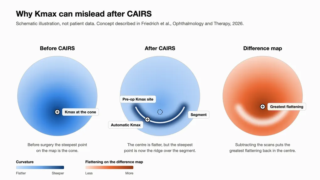
Kmax is the steepest measured curvature on the front of the cornea. It helps track keratoconus, but CAIRS changes the corneal surface and can change where that steepest point is measured.
That is the argument of Is Kmax a Reliable Endpoint for Evaluating Outcomes after CAIRS?, a commentary published in Ophthalmology and Therapy on 10 September 2026. It was led by Maximilian Friedrich and Hyeck-Soo Son at the University of Heidelberg, with co-authors from Pallas Kliniken in Switzerland, Dr Soosan Jacob of Dr Agarwal’s Eye Hospital in Chennai, who first described CAIRS in 2018, and Dr David Gunn of the Queensland Eye Institute.
What CAIRS does to the corneal surface
CAIRS places donor corneal tissue segments in the mid-periphery to flatten and regularise the centre. Local steepening can occur directly over a segment.
When Kmax moves onto the segment
A topographer finds Kmax by searching the map for its steepest point. Before surgery that is often near the cone. When CAIRS flattens the centre, the steepest point may shift to the ridge over the segment.
The commentary uses two illustrative eyes scanned with a Pentacam AXL Wave before CAIRS and at three months. Tangential Kmax rose in both despite central flattening, with the steepest point shifting towards the segment. Axial Kmax fell in one eye and rose in the other.
Measurements the authors propose
The authors propose measures that sit in the central cornea, where CAIRS is meant to act:
- mean keratometry, together with flat and steep keratometry
- the keratometry value at the exact location of the preoperative Kmax
- the greatest flattening on a difference map between the preoperative and postoperative scans
- Kmax calculated within the central 4 mm only, inside the inner edge of the channel
These measures are proposals, not validated replacements for detecting progression after CAIRS. The authors also ask that reports identify the topographer and curvature method.
Related findings in the Brisbane series
Dr Gunn’s 85-eye femtosecond CAIRS series, published in Clinical & Experimental Ophthalmology in 2026, showed a similar difference between Kmax and central corneal measurements. Mean keratometry fell from 49.5 D to 45.4 D and central 3 mm corneal power fell by 5.4 D. Kmax fell by 2.5 D on average and varied between eyes.
Cross-linking decisions after CAIRS
CAIRS has not been shown to stop keratoconus progression. Decisions about corneal cross-linking require serial scans read alongside vision, refraction and segment position. A changed Kmax alone cannot settle those decisions.
If you have had CAIRS
A scan report showing a higher Kmax than before surgery is not, by itself, a sign that your keratoconus has worsened. Bring the report to your next review so it can be read against your earlier scans and the position of your segments.
If you have keratoconus and want to know whether CAIRS suits your cornea, book an assessment. The commentary is open access at Ophthalmology and Therapy.
Frequently Asked Questions
What is Kmax?
Why can Kmax go up after CAIRS?
Does a higher Kmax after CAIRS mean the operation did not work?
What additional measurements do the authors propose?
How is keratoconus progression checked after CAIRS?
Is the commentary free to read?