Femtosecond CAIRS in 85 Eyes: The Brisbane Study and the Donor Tissue Question
On 13 March 2026, Clinical & Experimental Ophthalmology, the journal of the Royal Australian and New Zealand College of Ophthalmologists, published the outcomes of the Brisbane nomogram for femtosecond laser-created corneal allogenic intrastromal ring segments. The paper by Dr David Gunn, Dr Rebecca Cox and Dr Brendan Cronin of the Queensland Eye Institute appeared in the July print issue (volume 54, issue 5, pages 664–677) and is open access.
On 7 September the journal published a letter about the study from Elaine Chong and Elsie Chan, together with the authors’ reply. The letter questioned the use of donor tissue and the selection of patients for surgery.
What the study found
The retrospective, uncontrolled study followed 85 eyes of 75 patients with keratoconus, all unable to tolerate rigid contact lenses, for a mean of 7.5 months (range 3 to 32 months).
- Uncorrected vision improved by an average of 4 lines; the paper reports 31 eyes gaining 5 lines or more.
- Best corrected vision in glasses improved by an average of 2 lines, but 5 eyes lost corrected acuity: 4 lost one line and 1 lost two lines.
- Mean keratometry fell from 49.5 D to 45.4 D, and corneal power across the central 3 mm fell by 5.4 D.
- Total higher-order aberrations and vertical coma both fell (p < 0.001).
- No intraoperative or postoperative complications, including melt or segment extrusion, were recorded during follow-up.
Of the 85 eyes, 18 had been cross-linked before CAIRS, 30 had cross-linking at the same sitting, 10 had it at least three months later, and 27 had no sign of progression and no cross-linking. Eyes treated with simultaneous cross-linking had a greater fall in Kmax than eyes without it (−4.28 D against −0.70 D). Eyes cross-linked before CAIRS showed no significant difference in outcome from eyes that had not been, although the groups were small.
Limits of the study
The series had no control group and follow-up varied between eyes. With a mean follow-up of 7.5 months, it cannot establish long-term safety or how long the visual changes last. Cross-linking groups were not randomised, so their results cannot determine the best timing of cross-linking. The findings apply to the selected patients in the study, all of whom could not tolerate rigid lenses.
How the segments were made
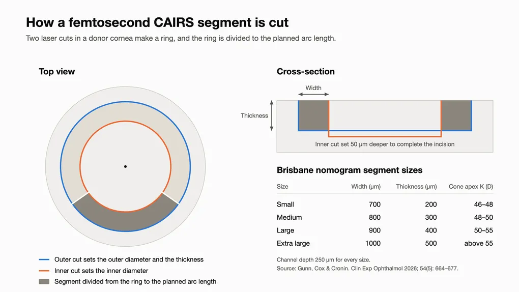
Every segment in the series was cut from a donor cornea on the WaveLight FS200 femtosecond laser, which also cut the channel in the patient’s eye. The laser makes two circular cuts in the donor tissue. The outer cut sets the outer diameter and the thickness of the ring, and the inner cut, set 50 µm deeper, sets its inner diameter. The ring is then measured and divided to the arc length in the plan.
The Brisbane nomogram uses four segment sizes, from 700 µm wide and 200 µm thick up to 1000 µm by 500 µm, chosen by the steepness of the cone. The channel is 250 µm deep for every size. Because the laser cuts the segment, its dimensions are known before it goes in, rather than depending on the thickness of the donor cornea. That is what allows outcomes to be measured against segment size, and the nomogram to be refined as results come in.
Donor tissue
Chong and Chan questioned the use of transplant-grade donor tissue for CAIRS. The segments in the series were cut from donor corneas that met Eye Bank Association of Australia and New Zealand viability standards, and donor corneas are a limited resource.
The reply agrees that donor tissue is scarce and explains the choice. When the Brisbane team began performing CAIRS in May 2021 there were no commercial segments available in Australia and very little published experience to draw on. Viable tissue was used so that tissue quality would not become another variable while the nomogram was being built. The endothelium is removed from every segment; CAIRS uses the stroma rather than the endothelial cells needed for an endothelial transplant.
Since that series closed, the authors’ reply describes a move to lamellar-grade donor corneas. A cornea with insufficient endothelial cells for an endothelial transplant can retain usable stroma, although donor screening and assessment for the intended use are still required. This does not mean every cornea unsuitable for one type of transplant is suitable for CAIRS. The reply describes measuring donor swelling with optical coherence tomography before cutting and adjusting cut depth to account for hydration. The authors also supported extending Medicare Benefits Schedule reimbursement to gamma-irradiated and pre-packaged segments.
Which patients should have CAIRS
The second question was patient selection, including whether eyes that already reach good acuity in glasses should be offered surgery. The reply argues against a threshold of 6/19 corrected vision.
In the study, 27 eyes had corrected vision of 6/7.5 or better before surgery. According to the reply, every one of those eyes needed at least 2 D of astigmatism corrected to reach that acuity, up to 7.5 D, and in each case the astigmatism was irregular. These patients had already failed rigid or mini-scleral lenses and reported poor quality of vision in both glasses and contact lenses. An acuity of 6/7.5 achieved through a correction the patient cannot wear is not functional vision.
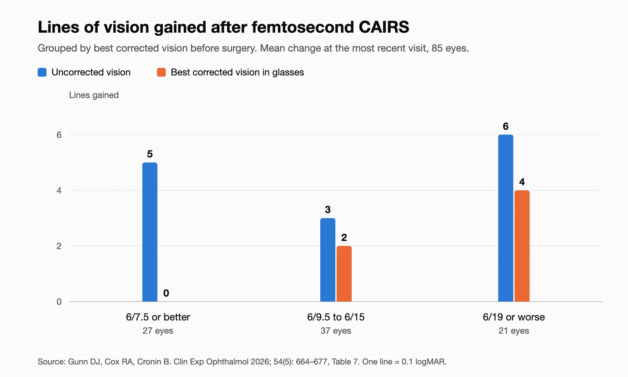
The study’s subgroup results describe the changes in those eyes. Their uncorrected vision improved by an average of 5 lines, while their best corrected acuity hardly changed, because it was already near the top of the chart. Eyes starting at 6/19 or worse gained the most on both measures. These subgroup observations do not predict how much an individual patient will improve.
Children and cross-linking
The authors’ reply also discusses CAIRS in children. The series included a range of ages and cannot establish the long-term benefits or risks specifically for children.
CAIRS is not proven to halt keratoconus progression. Where there is evidence of progression, cross-linking is considered separately. In the authors’ reply, Dr Gunn’s preference is to cross-link at a later date, because cross-linking produces some flattening of its own and staging the two lets the effect of each be assessed. The timing is decided for each patient.
Kmax also needs care when interpreting a scan after CAIRS. A separate 2026 commentary on Kmax after CAIRS discusses how local steepening over a segment can change that number despite central flattening.
Reading the papers
The study is open access at Clinical & Experimental Ophthalmology. The letter from Chong and Chan is at doi.org/10.1111/ceo.70167 and the reply at doi.org/10.1111/ceo.70166. Surgeons can plan cases with the Brisbane nomogram free at CAIRSPlan.
If you have keratoconus and your vision in glasses tests well in the clinic but does not work for you day to day, book an assessment to find out whether CAIRS suits your cornea.
Frequently Asked Questions
What is femtosecond CAIRS?
Does CAIRS use a cornea that could have gone to a transplant patient?
Can CAIRS help if my vision in glasses is already quite good?
Is cross-linking done at the same time as CAIRS?
Were there complications in the study?
Where can I read the study?