Accelerated Corneal Cross-Linking for Keratoconus in Children: A Queensland Children's Hospital Study
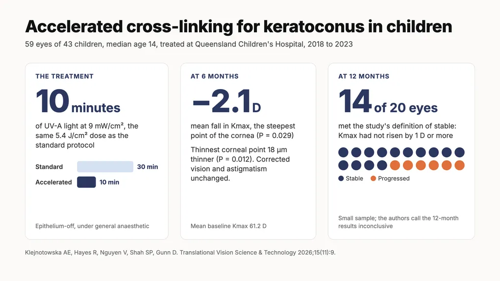
Keratoconus that starts in childhood tends to move faster than keratoconus that starts in adult life. A study published in Translational Vision Science & Technology in November 2026 reports what happened to 59 eyes of 43 children treated with accelerated corneal cross-linking at Queensland Children’s Hospital in Brisbane. Six months after treatment the steepest point of the cornea was, on average, 2.1 dioptres flatter.
The paper is Visual and Topographic Outcomes of Accelerated Corneal Cross-Linking for Keratoconus in Indigenous and Non-Indigenous Children in Australia, by Adrianna Klejnotowska, Rylan Hayes, Vivien Nguyen, Shaheen Shah and Dr David Gunn, from the hospital’s Department of Ophthalmology. Dr Gunn is the senior author. The authors state that it is the first study to report cross-linking outcomes for Aboriginal and Torres Strait Islander children.
Why keratoconus in children is treated early
The paper cites a prevalence of keratoconus as high as 1 in 84 among Australian 20-year-olds. In one series it quotes, 27.8% of patients younger than 15 already had advanced disease when first seen, against 7.8% of those aged 27 or older.
Cross-linking does not reverse keratoconus. It stiffens the cornea so the cone stops advancing, which is why it is offered as soon as scans show the cornea is changing. A child treated at 12 has decades of vision resting on that result.
The children in the study
The study reviewed every child who had accelerated cross-linking at the hospital between November 2018 and June 2023 and returned for at least six months of follow-up.
- 59 eyes of 43 children, median age 14 (range 9 to 17)
- 72.9% of eyes had advanced or severe keratoconus at baseline, with a mean Kmax of 61.2 D
- most children had recorded eye rubbing, allergic eye disease and an atopic background such as eczema, asthma or hay fever
- 9 of the 43 children (20.9%) were Aboriginal or Torres Strait Islander
Each eye was treated because Pentacam scans or vision testing showed progression over 6 to 12 months: a rise in Kmax of at least 1 D, a line of vision lost, astigmatism or short-sightedness up by at least 1 D, or the cornea thinning by more than 13 µm.
How accelerated cross-linking differs from the standard treatment
Standard cross-linking, the Dresden protocol, soaks the cornea in riboflavin (vitamin B2) and then exposes it to ultraviolet-A light at 3 mW/cm² for 30 minutes. Accelerated cross-linking delivers the same 5.4 J/cm² in a third of the time by using a beam three times as bright.
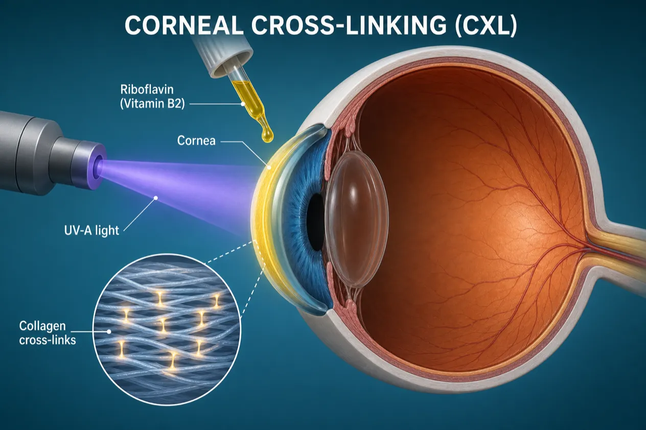
In the study every procedure was done under general anaesthetic. The surface layer of the cornea, the epithelium, was removed over the central 8 to 9 mm. Riboflavin 0.1% drops went on every 2 minutes for 20 minutes, the cornea was checked to be at least 400 µm thick, and UV-A light at 9 mW/cm² was applied for 10 minutes. A bandage contact lens was placed at the end, and antibiotic and steroid drops were used for two weeks.
Results at six months
Mean Kmax fell by 2.09 D (95% confidence interval 0.23 to 3.95, P = 0.029). The thinnest point of the cornea was 18 µm thinner (P = 0.012). Corrected vision and astigmatism did not change significantly.
Both changes held after the analysis adjusted for atopy, allergic eye disease and eye rubbing.
A thinner reading after cross-linking is a recognised finding, and it can look like progression on a scan report. The authors attribute it to the treated tissue becoming more compact and to remodelling of the epithelium. Their advice is to read corneal thickness after cross-linking together with the curvature maps, not by itself.
Results at 12 months
Twenty eyes had Kmax measured at both baseline and 12 months. Fourteen (70%) were stable. Six (30%) had steepened by 1 D or more and were classed as having progressed, a rate the authors describe as modestly higher than the 18% to 26% in two comparable published series. Corrected vision was 0.09 logMAR better on average, which was not statistically significant.
Much of the 12-month data is missing. COVID-19 restrictions turned many clinic visits into phone consultations, when no scan could be taken. The authors call the 12-month results inconclusive.
Outcomes for Aboriginal and Torres Strait Islander children
Aboriginal and Torres Strait Islander children made up 20.9% of the children in the study, against about 3.3% of the Australian population. The analysis found no significant association between Aboriginal and Torres Strait Islander status and any of the four outcomes at 6 or 12 months.
Two cautions come with that. Twelve eyes of nine children is a small group, so the study could miss a moderate difference. The over-representation may also reflect which children are referred to a public tertiary hospital. The authors call for research into the causes, risk factors and long-term outcomes of keratoconus in Aboriginal and Torres Strait Islander children.
Complications
Corneal haze was recorded in 22% of eyes, a sterile inflammatory infiltrate in 5.1% and infectious keratitis in 3.4%. All either settled without treatment or cleared with drops. Haze was graded at the slit lamp by the examining clinician, and the authors note that this is hard to measure objectively.
What the study cannot show
This was a retrospective review with no untreated group and no standard cross-linking group, so it cannot say how these eyes would have fared without treatment or whether the 10-minute protocol matches the 30-minute one. For that the paper points to the KERALINK randomised trial in young patients and to published comparisons of the two protocols, which found no significant difference in Kmax.
The paper’s section on future protocols cites the topography-guided, oxygen-supplemented epithelium-on technique that Dr Gunn co-authored with Dr Brendan Cronin in the Journal of Cataract & Refractive Surgery in 2024.
If your child has keratoconus
Keratoconus in a child or teenager is tracked with repeat corneal scans a few months apart. If the scans show change, cross-linking is usually recommended promptly. Glasses that need updating every few months and astigmatism that keeps rising are reasons to ask an optometrist for corneal topography.
Eye rubbing and allergic eye disease were recorded in most of the children in this study. Treating the itch so that the rubbing stops is part of managing the condition at any age.
To arrange an assessment with Dr Gunn, book an appointment; optometrists and GPs can refer online. The study is open access at Translational Vision Science & Technology.
Frequently Asked Questions
Can children have corneal cross-linking for keratoconus?
What is accelerated corneal cross-linking?
Does accelerated cross-linking work in children?
Is cross-linking done under general anaesthetic in children?
Do Aboriginal and Torres Strait Islander children respond differently to cross-linking?
What are the risks of cross-linking in children?
Is the study free to read?