Dr David Gunn at ESCRS 2026: Four CAIRS Talks in London
The 44th Congress of the European Society of Cataract and Refractive Surgeons ran from 11 to 15 September 2026 at ExCeL London, with the organisers expecting more than 15,000 delegates from over 130 countries. CAIRS appeared in two instructional courses, an industry symposium and four days of hands-on wet labs, and Dr David Gunn and Dr Brendan Cronin from the Queensland Eye Institute taught in all three. Dr Gunn had previewed the trip in August. His four talks, and what was said in each:
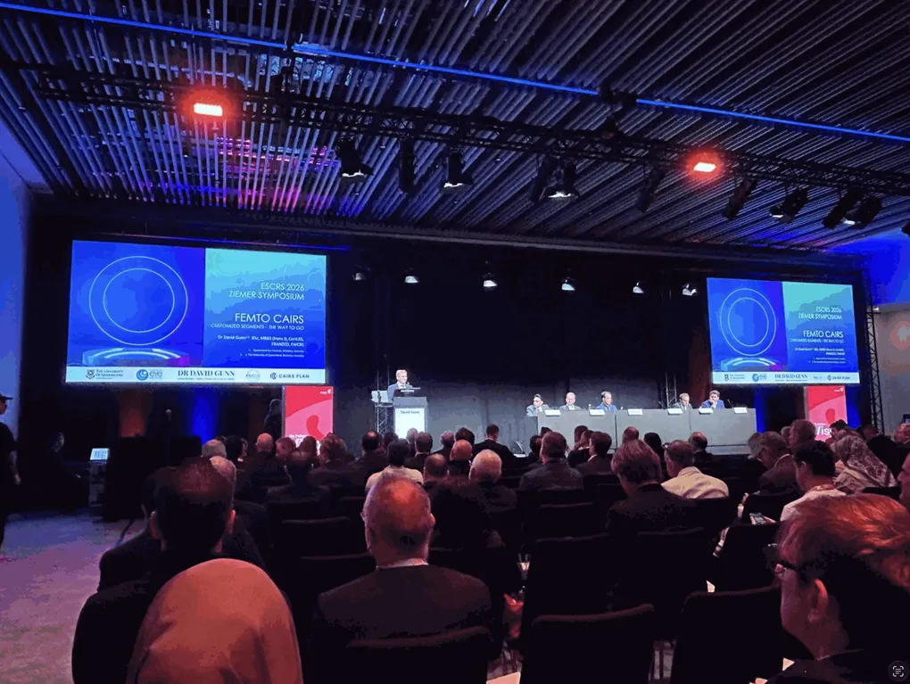
- Saturday 12 September, Ziemer satellite symposium: Femto CAIRS: Customised Segments, The Way to Go. Recorded by Ziemer.
- Monday 14 September, Transitioning to CAIRS, Dr Soosan Jacob’s instructional course: CAIRS by femtosecond laser.
- Monday 14 September, CAIRS in the Real World: Unplugged, Dr Hatch Mukherjee’s instructional course: Planning CAIRS cases and using online tools.
- Friday 11 to Monday 14 September, Lions World Vision Institute wet labs: the CAIRS wet lab and the CAIRS Planning and Resources workshop.
- Saturday 12 September, Ziemer podcast: Tips and tricks for starting CAIRS, recorded with Dr Brendan Cronin. Release pending.
Also at the congress, and covered at the end of this post, was the ePoster on early outcomes with the Clareon PanOptix Pro lens, presented with Dr Luisa H. Colorado of the Queensland University of Technology.
1. Ziemer symposium: Femto CAIRS with customised segments
Ziemer satellite symposium, Saturday 12 September, 13:00 to 14:00, George V Room 3. Ziemer’s lunchtime session, Redefining the Future: We Have It All, was chaired by Professor Jod Mehta of Singapore and ran the FEMTO LDV Z8 across its uses: Professor Theo Seiler on femtosecond cataract surgery, Dr Florian Bauer on posterior capsulotomy, Dr Gunn on Femto CAIRS, Professor Shady Awwad on centration for CLEAR Supra with FlowSuite, Nicolas Lutrand on the AQUARIUZ ablation platform, and Professor Mehta on transepithelial femtorefractive keratectomy.
Ziemer filmed the whole session. It is on YouTube as ESCRS Symposium 2026, 56 minutes, and Dr Gunn’s talk starts at 16:36 and runs ten minutes.
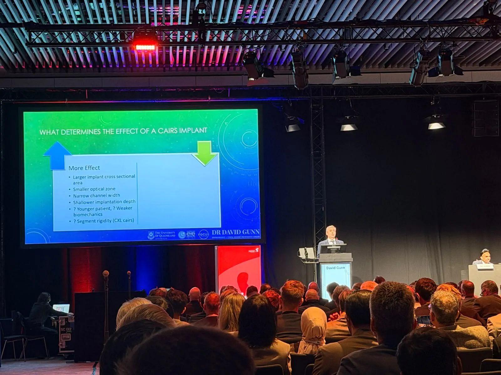
What was said. The talk opened on one question: what determines the effect of a CAIRS implant? More effect comes from a larger implant cross-sectional area, a smaller optical zone, a narrower channel and a shallower implantation depth. A younger patient or a weaker cornea probably adds to it, and a cross-linked segment may behave differently again. Every one of those is a number the surgeon chooses or measures, which is the whole case for planning the segment rather than picking one off a shelf.
From there it followed Dr Gunn’s own route to the platform. Dr Bader Khayat performed the first femtosecond CAIRS; Dr Gunn was the second surgeon to do so and the first on the WaveLight FS200, in 2021, and he performed the first Z8 CAIRS in Australia in 2024. The Z8 software cuts a segment symmetric, tapered or double-tapered, with its position adjusted live against the marks on the eye, so two segments in the same cornea can be different shapes when the cone is asymmetric. The Brisbane nomogram, refined from 85 published eyes and two further years of outcomes, turns those options into a plan. The talk then worked through two cases, a severe cone and a mild one, and the paired unequal segments that customised cutting makes possible.

Straight after the symposium, Dr Gunn and Dr Cronin recorded a podcast for Ziemer in a studio set up at the venue: Tips and Tricks for Starting CAIRS, a conversation for surgeons taking on the procedure, from planning the first case to preparing and placing the segment. Ziemer’s highlights reel from the second day carried a clip; the episode is Ziemer’s to release, and this post will link it when it is out.
2. Transitioning to CAIRS, Dr Soosan Jacob’s course: the femtosecond route
Transitioning to CAIRS, ESCRS instructional course, Monday 14 September. Led by Dr Soosan Jacob of Chennai, who devised the technique, and pitched at surgeons starting out. Ten-minute talks: Dr Jacob on what CAIRS is and what is needed to start, Dr Shady Awwad (Beirut) on results and safety, Dr Bader Khayat (Munich) on surgical steps, then Dr Gunn on making the segment with a femtosecond laser and Dr Cronin on making it with a trephine, placed back to back so the room could weigh the two routes.
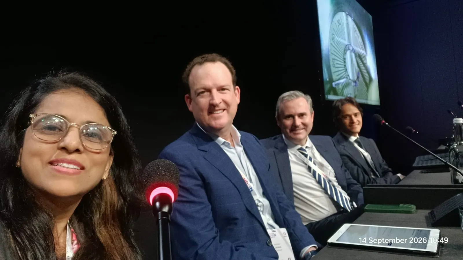
What was said. Dr Gunn’s ten minutes started where a surgeon starting out actually starts, with the decision about how the donor tissue will be cut. There are four ways to make a segment, and he set out the drawbacks of each, including his own. Your own trephines and a donor cornea: cheap and fast, but the thickness of the tissue and the exactness of the second cut are uncertain. A purpose-made double-bladed trephine: more accurate, but fixed sizes, and still no measure of thickness. Precut, sterilised segments from an eye bank: ready to use with a long shelf life, harder to shape finely, and variable in thickness. Segments cut on a femtosecond laser: full control of thickness, width and arc, at the cost of a donor interface per case, a laser in the day hospital, five to ten minutes of cutting and more technique to learn.
The argument for the fourth is not that it is easier. A segment with known dimensions can be measured against its outcome, and only then can a nomogram be built and refined. The Brisbane series of femtosecond-cut segments, 85 eyes of 75 patients with 43.7 per cent gaining five or more lines of unaided distance vision, was published with Dr Rebecca Cox and Dr Cronin in Clinical & Experimental Ophthalmology in 2026 (DOI 10.1111/ceo.70104). One slide is worth repeating for anyone starting: the difference between a segment’s radius and its diameter. Not every nomogram, laser interface and order form uses the same convention, and a segment specified on the wrong one is out by a factor of two.
3. CAIRS in the Real World, Dr Hatch Mukherjee’s course: planning cases and online tools
CAIRS in the Real World: Unplugged, ESCRS instructional course, Monday 14 September. A new course for 2026, led by Dr Achyut (Hatch) Mukherjee of Colchester and pitched at surgeons already performing CAIRS, with Dr Muhammad Hamza (Southampton) on reading topography, Dr Bader Khayat on insertion, Dr Rajesh Fogla (Hyderabad) on manual versus femto, Dr Farida Lafdjah (Doha), and Dr Mukherjee closing on troubleshooting and rescue: under-effect, over-effect, malposition, slit-lamp adjustment, split segments and extrusion.
What was said. Dr Fogla had just argued manual versus femtosecond, so Dr Gunn started one step downstream: given that you have chosen your method, how do you decide what to cut and where to put it? The workflow, in order: where is the cornea steep; one segment or two; how much tissue does the cone need, read from the topography on an absolute colour scale; and only then which dimensions and which arc, the decision surgeons new to the technique tend to start with. The Brisbane nomogram table was shown in full, along with the tapered and paired-unequal-segment cases that customised cutting makes possible.

The tool that runs that workflow is CAIRSPlan, which Dr Gunn co-developed with Dr Cronin and declared as his interest at the start of the talk. CAIRSPlan 2.0, released in May 2026 after eighteen months of use by 143 surgeons on the original tool, standardises every topography export onto one 201 by 201 grid at 0.05 mm before planning, strips patient identifiers at import, offers three nomograms (Brisbane 2026, Istanbul and Awwad), proposes a starting plan from the cone pattern with one click, and lets each segment be given custom width and thickness at three control points, which is what the Z8 then cuts. It is free, and by late September 2026 about 380 surgeons were using it. A five-minute walkthrough, from scan to surgical plan, is on Dr Gunn’s YouTube channel.
4. Lions World Vision Institute wet labs
LWVI stand, Friday 11 to Monday 14 September. The Lions World Vision Institute, the Tampa eye bank that supplies OptiGraft precut CAIRS segments, ran CAIRS, DSAEK and DMEK wet labs on the exhibition floor with an international instructor group that included Dr Jacob and both Brisbane surgeons. On the Friday Dr Cronin presented the CAIRS planning session with CAIRSplan.com at 13:15, then Dr Gunn led the CAIRS wet lab from 14:00 to 15:30 with Dr Cronin co-instructing, and both returned over the following days for further sessions, including the CAIRS Planning and Resources workshop.
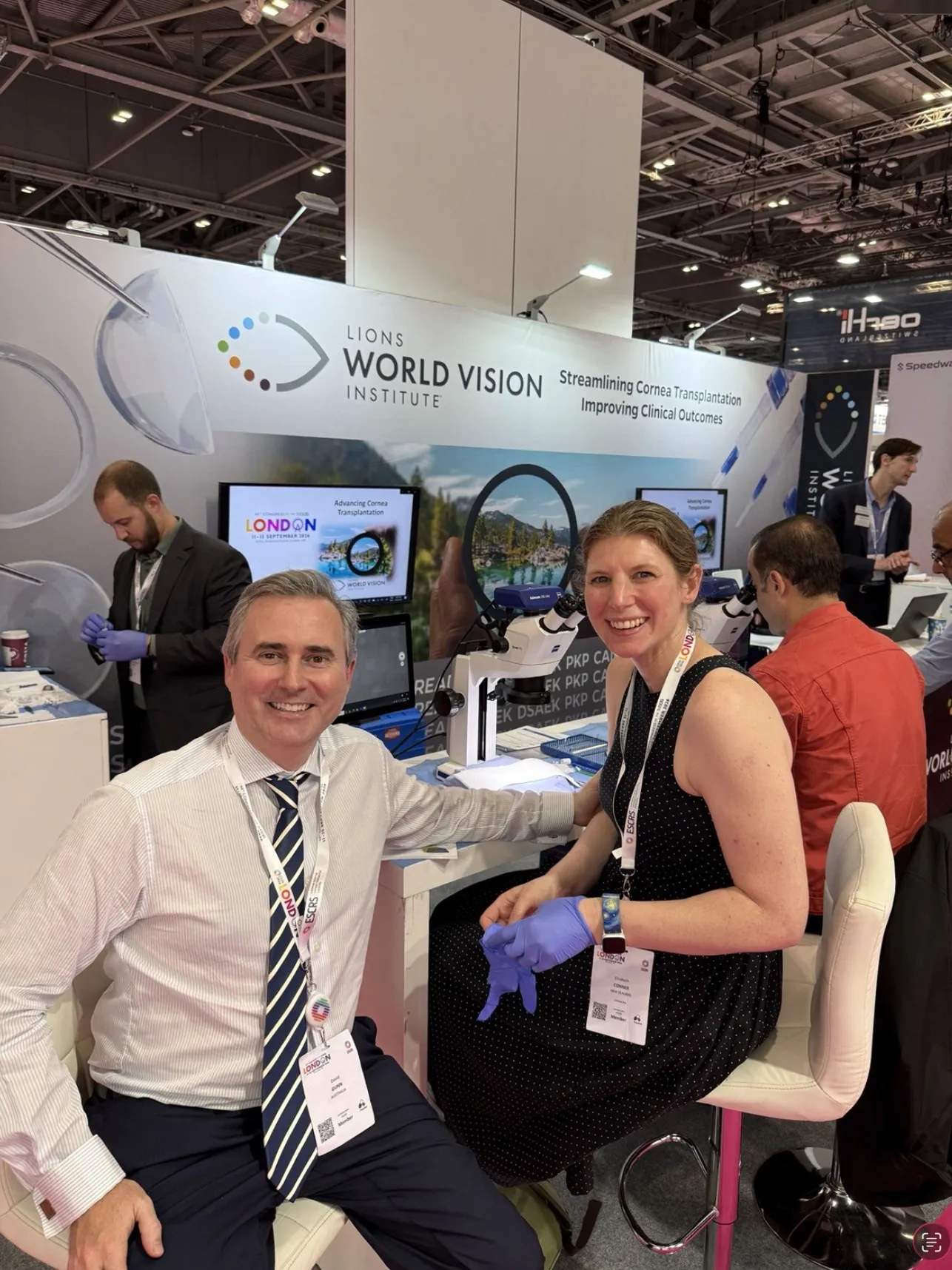
What was taught. The steps that decide a CAIRS result are small and physical. Handling donor tissue without folding it. Preparing and drying the segment, Bowman’s layer side down on a warmed surface for a timed interval, which is Dr Shady Awwad’s technique and is credited to him every time it is shown. The three channel numbers, depth, inner diameter and arc, and what each looks like under the microscope. Feeding the segment along the channel without hydrating it mid-way, and centring it on the marks rather than the pupil. And the plan the dimensions come from, so that surgeons left knowing not only how to place a segment but how to choose one.
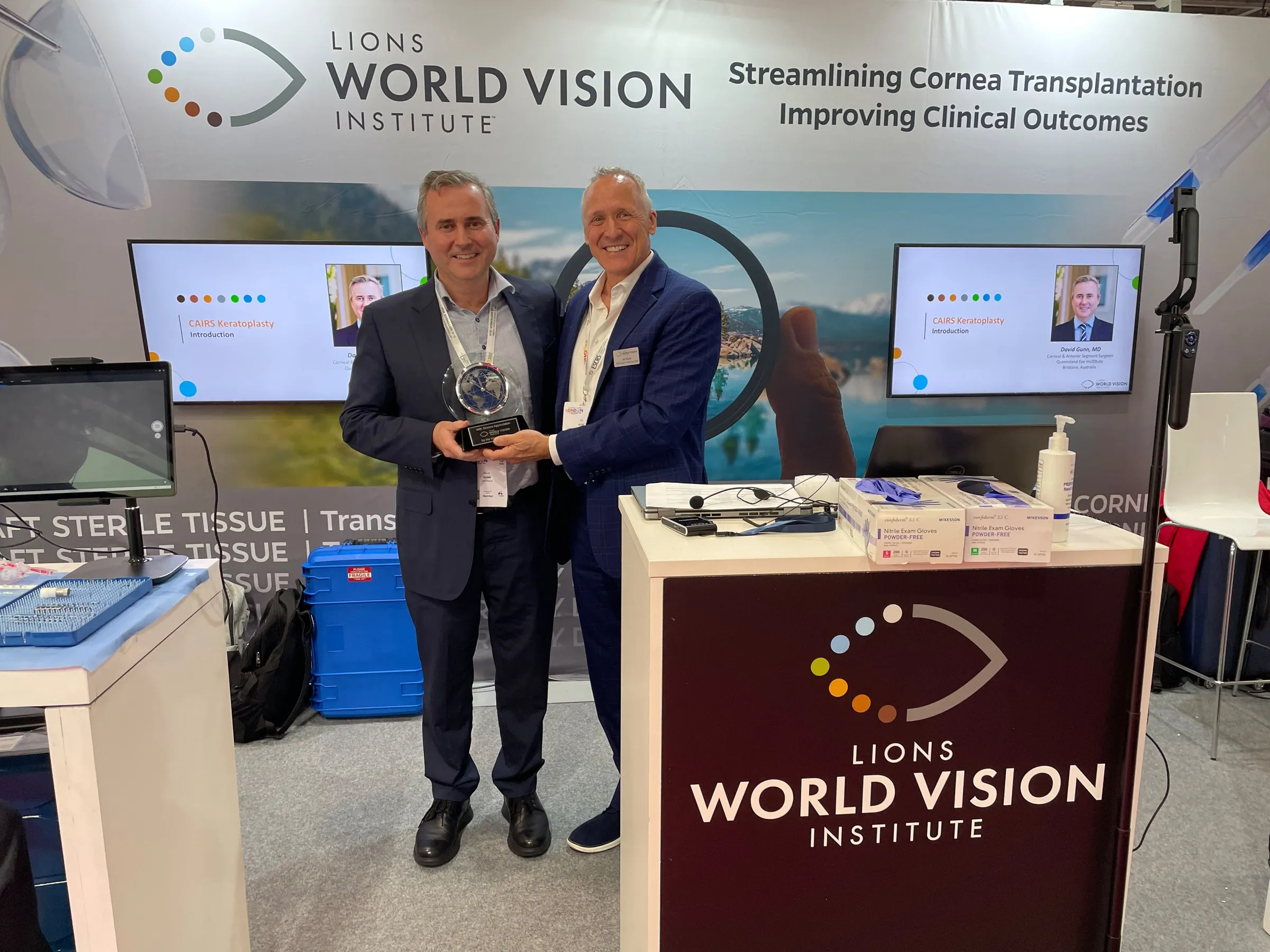
On the Sunday, LWVI presented Dr Gunn with a glass clock at its stand.
Also at the congress: the PanOptix Pro ePoster
Away from the cornea programme, Dr Gunn and Dr Luisa H. Colorado of the Queensland University of Technology presented an ePoster and presented poster, POCAT484, titled Early Real-World Visual and Patient-Reported Outcomes Following Bilateral Implantation of the Clareon PanOptix Pro Intraocular Lens. It is the interim analysis of an investigator-initiated, prospective, single-centre study of bilateral Clareon PanOptix Pro implantation targeting emmetropia, planned for 50 patients and reporting the first 11 patients, 22 eyes, at three months.
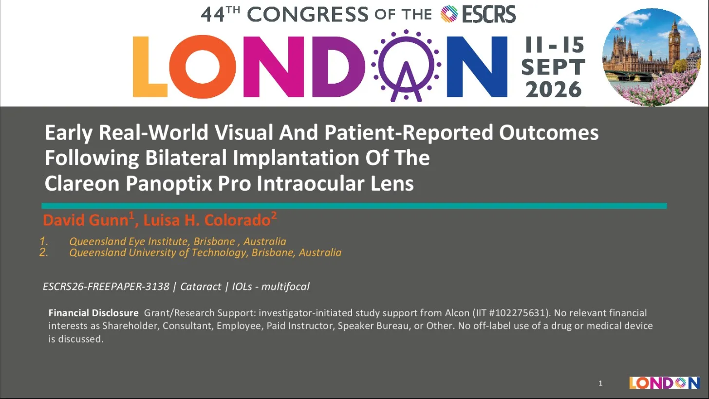
At three months, mean spherical equivalent had moved from +0.95 D before surgery to +0.16 D, with 91 per cent of eyes within 0.50 D of plano. Mean binocular unaided acuity was -0.01 logMAR at distance and at 66 cm and 0.04 logMAR at 40 cm, and binocular distance-corrected acuity stayed at 0.20 logMAR or better across the whole defocus range from +1.00 D to -3.00 D. On the IOLSAT and QUVID questionnaires, all eleven patients reported that they did not need glasses, would choose the same lens again and would recommend it; mean overall satisfaction was 3.8 out of 4, no visual symptom was rated “very much” bothersome, and ten of the eleven would not want further surgery for symptoms. The authors’ conclusion is a cautious one: the early results support the lens’s visual performance and patient acceptance and sit in line with published real-world outcomes for the first-generation Clareon PanOptix, but this is a single-centre interim dataset of 11 of 50 planned patients, and recruitment and follow-up continue. The study is supported by Alcon as an investigator-initiated trial (IIT 102275631), which is declared on the poster. ESCRS publishes accepted abstracts with a DOI in its Abstract Repository after each congress; the London 2026 set was not yet online when this was written, and the entry will be linked from the publications list when it is.
Credits and disclosure
Dr Soosan Jacob devised CAIRS, and every talk on the subject starts there. Dr Shady Awwad developed the clear marker used for centration, the segment drying technique and the mould used in theatre, and worked with Dr Gunn and Ziemer on the all-femtosecond CAIRS modules. Dr Gunn thanks Pascal Eberhard, Alba Segura and the Ziemer team for the symposium and the podcast, Jeremy Shuman and LWVI for the wet labs, and Dr Brendan Cronin for the week.
Dr Gunn has a commercial relationship with Ziemer: he speaks at the company’s meetings and provides clinical feedback on the Z8 CAIRS software, and he owns the laser he uses. He co-developed CAIRSPlan. Neither changes the numbers above, which come from the published Brisbane series.
For patients in Brisbane
Everything presented in London is the operation performed here. Dr Gunn performs femtosecond CAIRS at the Queensland Eye Institute for keratoconus that has stabilised after cross-linking but still limits vision in glasses or contact lenses. Book an assessment to find out whether your cornea suits it.
Frequently Asked Questions
What is CAIRS?
Where can I watch Dr Gunn's ESCRS 2026 talk?
What is femtosecond CAIRS with customised segments?
What is CAIRSPlan?
What is the Lions World Vision Institute?
What did the PanOptix Pro ePoster report?
Is femtosecond CAIRS available in Brisbane?