Keratoconus at AUSCRS 2026: Chairing Cornea Saturday Morning Live, The Cone Edition
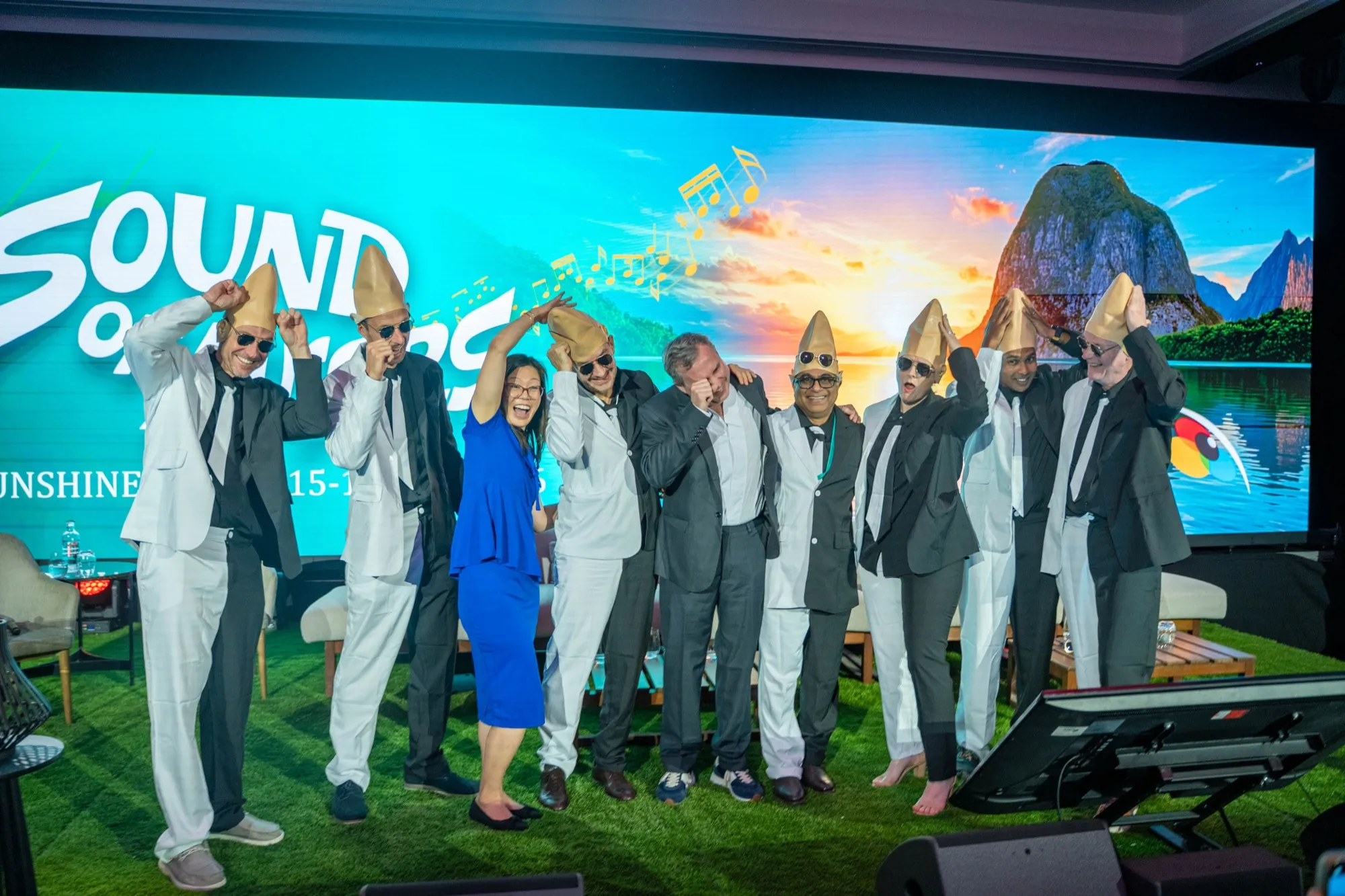
AUSCRS held its 2026 meeting at the Novotel Sunshine Coast at Twin Waters from 15 to 18 July, under the title The Sound of AUSCRS. The cornea session closed the scientific programme on the Saturday morning, and it was not run as a lecture.
A cornea session built as a talk show
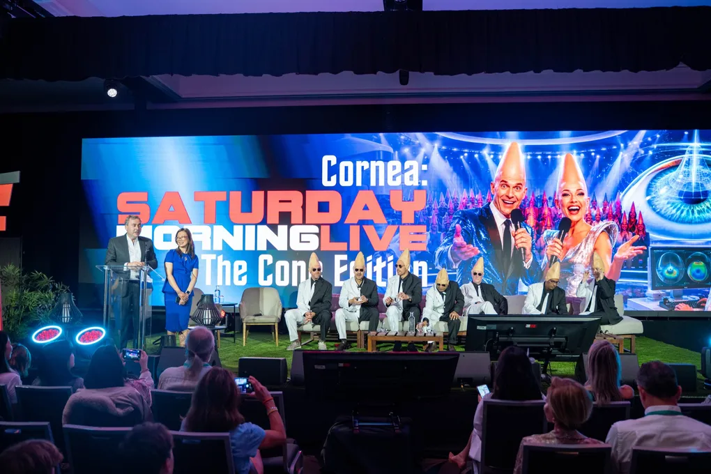
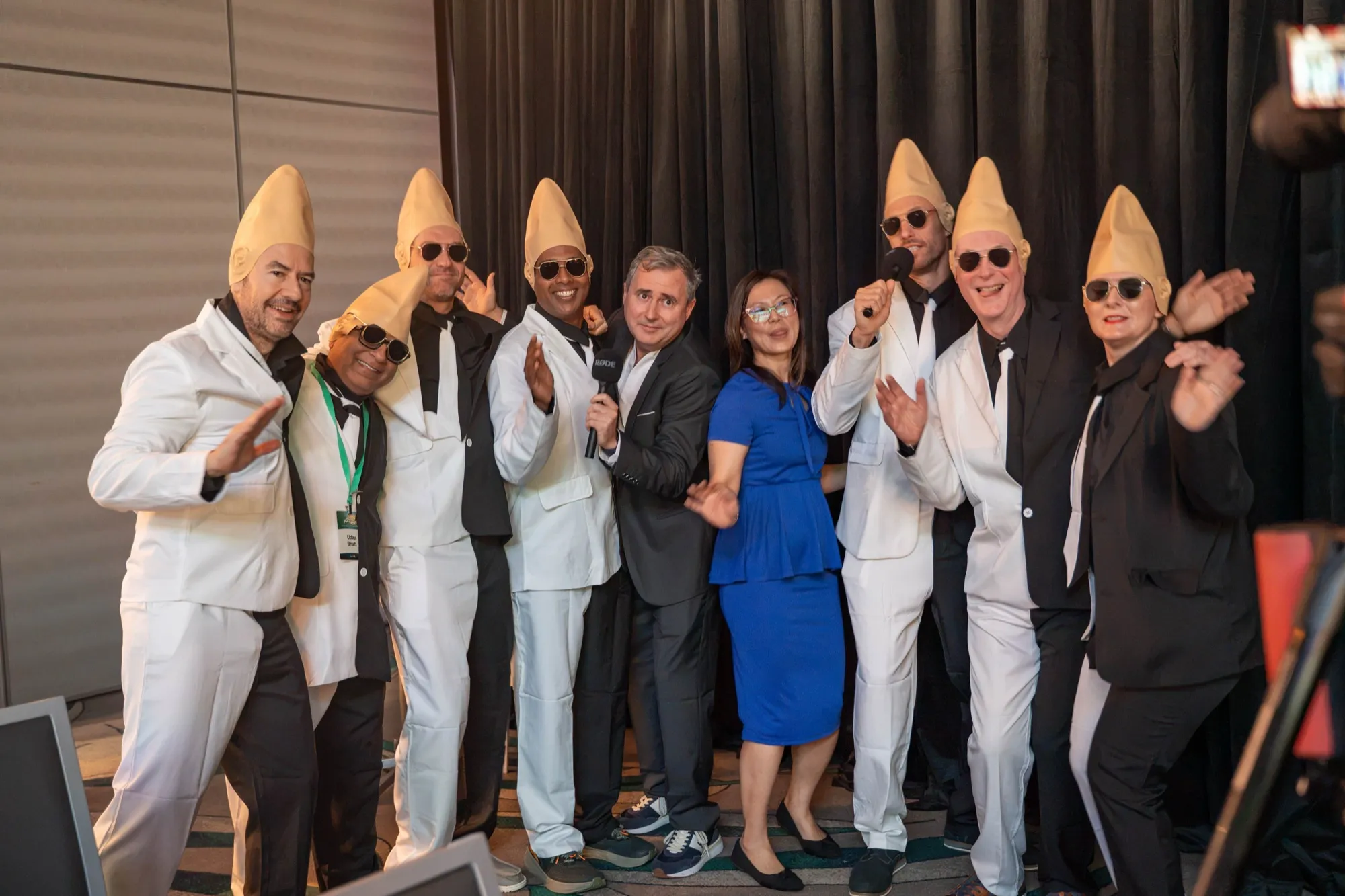
Session Ten, Cornea: Saturday Morning Live — The Cone Edition, ran from 10.30 to 12.00 on Saturday 18 July. Dr David Gunn co-chaired it with Dr Aanchal Gupta and presented in it. The set was built as a live television studio, with sofas, lawn and a title sequence, and the panel appeared in Coneheads costume.
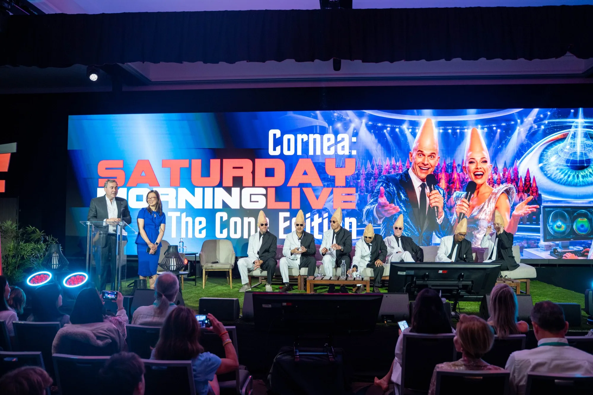
The costume is a pun on the disease. It also solved a real scheduling problem. Cornea is usually the last session on the last morning of a cataract and refractive meeting, and a Saturday slot after four days is where attention normally goes to die. The room was full.
Who spoke, and what they argued
Eight talks ran in ninety minutes, which is the format’s discipline. Nobody gets long enough to be dull.
Professor Damien Gatinel, head of anterior segment and refractive surgery at the Rothschild Foundation in Paris, opened the causation argument with Stop Rubbing Your Eyes! Establishing Eye Rubbing as a Cause of Keratoconus: A Bradford Hill Demonstration — The Vegemite Way. Bradford Hill’s criteria are the standard epidemiological test for deciding whether an association is causal rather than coincidental, and Professor Gatinel’s position, set out at length on his own site and in the literature as the no rub, no cone conjecture, is that eye rubbing satisfies them. The claim is still contested and the session did not settle it. It does change what a consultation is for: if rubbing is causal, then asking about it and stopping it is treatment, not advice.
Dr Dylan Joseph, a cataract and refractive surgeon in Knysna, South Africa, presented READ Laser Treatment and Allotex Corneal Inlays — Corneal Solutions for Presbyopia. Allotex inlays are donor corneal tissue shaped by laser and placed in the cornea to restore near vision, which is the same idea as CAIRS pointed at a different problem: allogenic tissue, cut by laser, used to change the shape of the cornea without putting plastic in it. Dr Joseph trained in refractive and lens surgery in Ireland under Arthur Cummings and was part of the first clinical trials of the Streamlight procedure.
Mr Kieren Darcy argued for going beyond shape stabilisation in keratoectasia with a staged management plan aimed at unaided quality of vision, which is the same gap the rest of the session kept returning to. Professor Michael Goggin presented raytracing-driven corneal remodelling with the iVis excimer platform in stable keratoconus, and Dr Uday Bhatt compared untreated keratoconus progression across age groups. On the cataract side of cornea, Dr Georgia Cleary examined K versus TK for intraocular lens power calculation in eyes with mild corneal endothelial disease, and Associate Professor Chameen Samarawickrama covered the IC-8 lens in highly abnormal corneas.
Dr Gunn’s own talk, A Few of My Favourite ‘Rings’: CAIRS Plan Pro, was about the planning software rather than the surgery. The meeting’s Sound of Music theme reached the titles, which tells you something about the room.
What the cornea room is arguing about in 2026
Two questions took up most of the session, and neither is settled.
The first is how to cross-link. Corneal cross-linking has been standard treatment for progressive keratoconus for well over a decade, but the protocol keeps moving. The original Dresden technique removed the epithelium and left patients in pain for three to five days. Epithelium-on protocols avoid that, at the cost of oxygen, which is the rate-limiting ingredient in the reaction. Supplemental oxygen through a sealed goggle and pulsed ultraviolet-A delivery are the two modifications that recover the difference.
That is not a theoretical position on Dr Gunn’s part. The Queensland Eye Institute series of oxygen-supplemented, topography-guided epithelium-on cross-linking that he co-authored with Dr Brendan Cronin (Cronin, Gunn, Chang and colleagues, Journal of Cataract and Refractive Surgery, 2024) covered 102 eyes over a mean of 11.5 months, with significant improvement in corrected distance visual acuity, average keratometry and maximum keratometry, and no complications (PubMed).
The second question is what to do about the vision once the cornea has stopped changing. Cross-linking arrests progression. It does not reliably give back the acuity a cone has already cost, and a patient whose keratoconus is stable but who still cannot function in contact lenses has a problem that stabilisation does not solve.
Ring segments, and how the channel is made
CAIRS puts allogenic corneal tissue into a channel in the recipient’s own stroma to flatten the cone and rebalance the cornea around it. Dr Gunn performed the first Australian case in May 2021 at the Queensland Eye Institute with Dr Brendan Cronin.
The channel is the enabling step, and it can be made by hand or with a femtosecond laser. Nearly everyone doing CAIRS now uses the laser, Dr Gunn included, because it fixes depth, diameter and arc as numbers rather than leaving them to the surgeon’s feel on the day. That part is no longer unusual.
The segment is where this practice differs. Dr Gunn cuts the segment with the laser too, so the dimensions of the implant are known before it goes in rather than approximated, and he was the second surgeon in the world to perform CAIRS that way. He was the first to do it on the WaveLight FS200 and on the VisuMax 800. Newer Ziemer Z8 software goes further again and allows the shape of each segment to be customised on the laser, so a segment can be cut differently for every cornea it is going into. Dr Gunn was the second surgeon in the world to have access to it and remains one of only a handful using it.
Reproducibility is what makes a nomogram possible, and without a nomogram every surgeon builds their own experience from scratch and nothing generalises. The planning tool Dr Gunn and Dr Cronin built at CAIRSPlan exists because the channel became a parameter rather than a manoeuvre.
The five-year Australian experience with femtosecond-created segments was published by Gunn, Cox and Cronin in Clinical and Experimental Ophthalmology in March 2026 (DOI 10.1111/ceo.70104).
The cases where segments are the wrong answer
Very steep, very thin corneas run past the range segments can correct, and scarring through the visual axis is not a shape problem. Those eyes still need a graft, and a session that only discussed ring segments would have left that out.
This is the least glamorous part of a keratoconus practice and the part patients most need explained. Ring segments are reversible and tissue-sparing, which is why they are tried first where the numbers support them. When the numbers do not, a corneal transplant is a better operation, not a failure of the smaller one.
For patients in Brisbane
Everything discussed on the Sunshine Coast is performed here. Cross-linking, femtosecond CAIRS and corneal transplantation are all done in Brisbane, and the assessment that decides between them is the same tomography and topography that the session spent two hours arguing over.
If keratoconus has been diagnosed and the next step is unclear, or if cross-linking has stabilised the cornea without restoring useful vision, book an assessment and the options can be mapped against the actual shape of the cone.
Frequently Asked Questions
What is AUSCRS?
What is a cone in keratoconus?
Does cross-linking improve vision, or only stop the keratoconus getting worse?
How long has CAIRS been performed in Australia?
Is CAIRS suitable for every cone?
Can CAIRS be combined with cross-linking?
Where can a patient in Brisbane be assessed for keratoconus surgery?