Topography-Guided PTK With Cross-Linking for Keratoconus at ESCRS Milan 2022
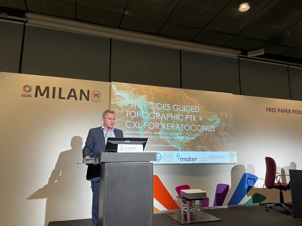
The 40th Congress of the ESCRS ran from 16 to 20 September 2022 at the Allianz MiCo in Milan. It was the society’s first congress back at full scale after the pandemic years, and the corridors felt like it.
Dr David Gunn had two pieces of work in the programme: a free paper with Dr Brendan Cronin on topography-guided phototherapeutic keratectomy combined with cross-linking, and a presented poster on a technique the Brisbane group had used in a patient nobody had published on before.
The free paper: reshaping and strengthening in one sitting
The free paper was titled Phorcides Guided Topographic PTK + CXL for Keratoconus, from the Queensland Eye Institute and Mater Hospital Brisbane.
The clinical problem is familiar to anyone who manages keratoconus. Cross-linking reliably stops the disease progressing, and that alone is worth doing. What it does not do is make the cornea a better optical surface. Patients whose keratoconus has been halted are often still stuck with a cornea too irregular for glasses and only tolerable in a rigid contact lens.
Topography-guided surface laser addresses the other half. The Phorcides Analytic Engine takes tomography data and works out a treatment that reduces the irregularity rather than chasing the spectacle prescription. Applied as a shallow therapeutic ablation and paired with cross-linking in the same session, the cornea comes out both more regular and structurally stronger.
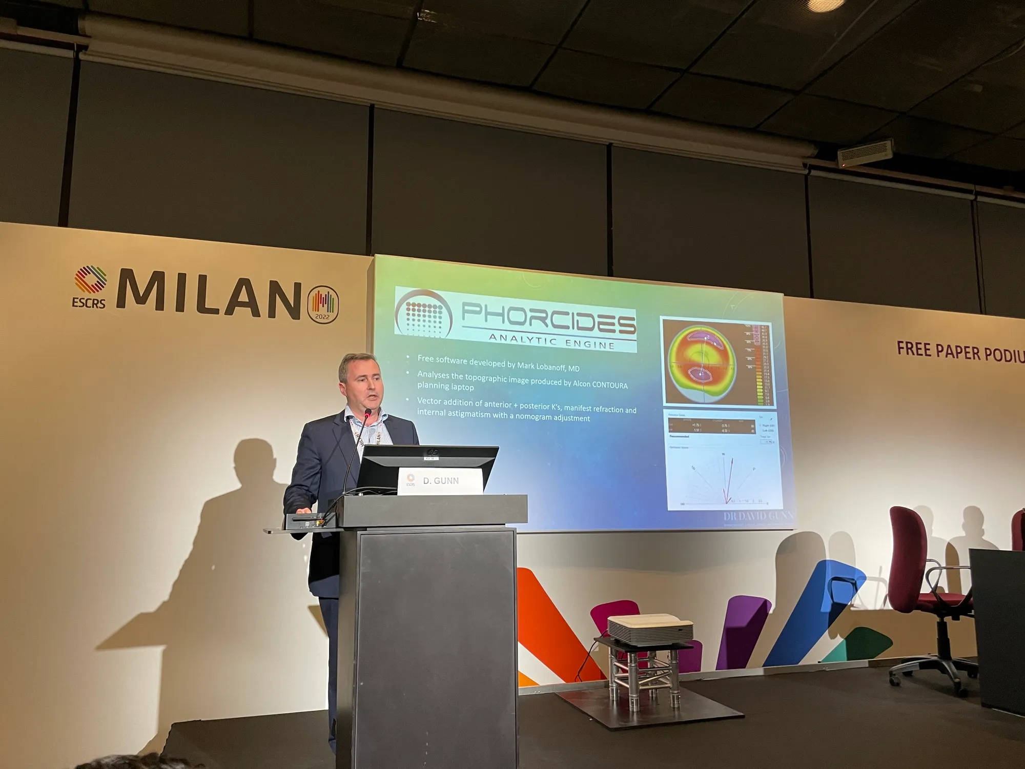
The discipline in this operation is restraint. Every micron of tissue you remove is a micron you cannot put back, and these corneas start thin. Dr Gunn plans to a tissue budget first and accepts a partial optical improvement inside it. Patients are told plainly that the goal is a cornea that works better in glasses or a soft lens, not a cornea that looks normal on a map.
The poster: CAIRS and cross-linking together after LASIK
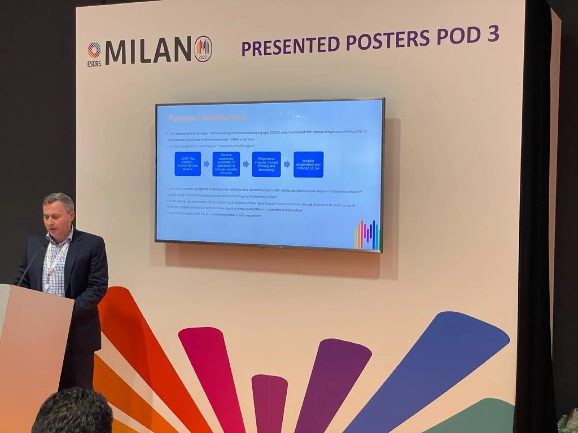
The second presentation, in the Presented Posters session, was a case report of CAIRS combined with cross-linking for post-LASIK keratectasia. As far as could be established at the time, it was the first report of that combination.
Post-LASIK ectasia is an unforgiving problem. The cornea has already had tissue removed, so the laser options that help in virgin keratoconus are largely closed off. Corneal allogenic intrastromal ring segments sidestep that, because they add donor corneal tissue into a channel rather than subtracting anything. Cross-linking in the same setting deals with the underlying weakness.
One case is one case, and the report said so. It opened a line of work that is now a routine part of the Brisbane practice for ectasia after refractive surgery.
Why the sequence matters more than the procedure
Both presentations came from the same position: these corneas need a plan rather than a procedure. A patient with progressive keratoconus, a distorted surface and contact lens intolerance may need cross-linking, laser regularisation and ring segments at different points, and the sequence matters.
Milan was where that argument first went to an international audience. The questions afterwards changed how the Brisbane group selected patients over the following year, which is the point of presenting rather than only publishing.
If you have been told you have keratoconus or ectasia after laser surgery, a consultation starts with tomography and a conversation about which of these tools applies to your cornea, and in what order.
Frequently Asked Questions
What is topography-guided PTK for keratoconus?
Why combine laser reshaping with cross-linking?
How much tissue is removed?
Is this suitable for every patient with keratoconus?