
Dr Gunn presents CAIRS for the prestigious Ridley Lecture at the German Ophthalmic Society meeting DOC May 2025

Dr David Gunn delivering the Ridley Lecture at DOC 2025 in Nuremberg
Honoured to Deliver the Ridley Lecture at DOC 2025, Nuremberg
I was deeply privileged to travel to Nürnberg Messe this month for the 37th International Congress of German Ophthalmic Surgery (DOC 2025), held 15–17 May at the NCC Ost convention centre in Nuremberg, Germany. Widely regarded as Europe’s premier cataract, corneal and refractive surgery meeting, DOC brought together more than 6,700 surgeons, researchers and industry innovators for three days of live surgery, plenary debates and hands-on training.
Presenting the Ridley Lecture — “CAIRS: A Paradigm Shift in Corneal Ectasia Management”
The highlight of my visit was the honour of giving the Harold Ridley Memorial Lecture in the General Session on Friday 16 May. My talk, “CAIRS – a paradigm shift in the management of corneal ectasia,” explored how Corneal Allogenic Intrastromal Ring Segments (CAIRS) are redefining treatment algorithms for progressive keratoconus and post-LASIK ectasia. I reviewed long-term biomechanical data, shared the latest nomogram refinements developed through the Brisbane nomogram and advancew in out planning software CairsPlan.com, and presented outcomes that show significant gains in corneal symmetry and uncorrected visual acuity.
Delivering the Ridley Lecture — named for Sir Harold Ridley, pioneer of the intra-ocular lens — was a career milestone. I was grateful for the opportunity to showcase Australian innovation on a global stage and to highlight how CAIRS complements cross-linking, topography-guided PRK and deep anterior lamellar keratoplasty in a modern keratoconus service.
CAIRS Experts Session
Prof Gerd Auffarth organised a experts CAIRS course with the surgeons giving their different perspectives on CAIRS. This group included some of the key pioneers in the technqiue Dr Aylin Kilic from Turkey and Dr Bader Khayat from Germany amongst other eminent ophthalmologists.


Gratitude to the DOC Leadership
My sincere thanks go to Prof. Dr. Michael C. Knorz (Mannheim) and Prof. Dr. Armin Scharrer (Fürth), for the invitation to speak and whose stewardship continues to make DOC an unmissable fixture on the international ophthalmic calendar. Their impeccable organisation created an atmosphere where rigorous science, practical pearls and collegial exchange thrived.
Key Take-aways for Colleagues and Patients
Global appetite for CAIRS is accelerating. Surgeons from Europe, the Middle East and Asia reported rapid uptake as an intermediate step to avoid keratoplasty for keratoconus patients.
AI and sustainability dominated the 2025 programme. Sessions on predictive analytics for IOL power and net-zero operating theatres underscored where our speciality is heading.
Interdisciplinary conversation is essential. From paediatric retina to cornea biomechanics, DOC reminded us that collaborative science speeds translational breakthroughs.
Keratoconus Background
Keratoconus is a disease of the cornea - the clear circular window at the front of the eye. The cornea normally has a regular spherical shape like a soccer ball. In keratoconus the cornea degenerates and becomes shaped more like a cone. This causes blurred and distorted vision. In the early stages of keratoconus an optometrist can fit glasses or contact lenses to help. Corneal cross linking is used to stop the disease from getting worse but has a very limited effect on improving the vision. Read more about Keratoconus and CAIRS treatment.
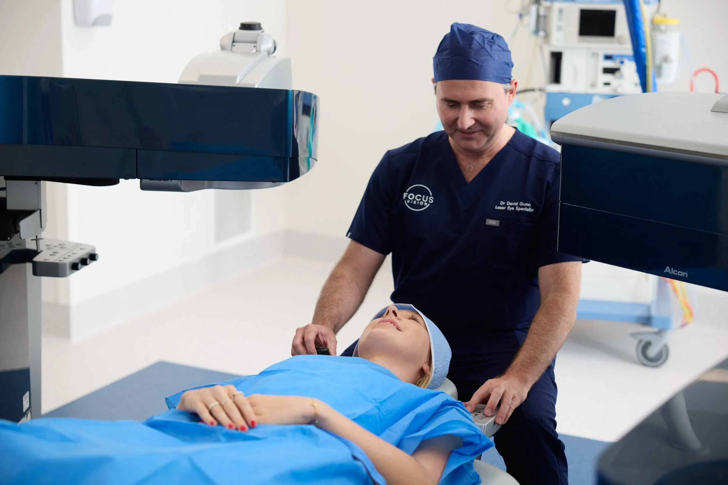
The first Corneal Allogenic Intrastromal Ring Segment (CAIRS) transplant surgeries in Australia were performed at the Queensland Eye Institute in May 2021 by Dr David Gunn and Dr Brendan Cronin, paving the way to better sight for many sufferers of the relatively common condition called keratoconus.

BENEFITS OF CAIRS
Human donor cornea tissue has a large number of benefits over plastic implants.
Biocompatibility
The main benefit of using corneal tissue is that it is a “like into like” implant. Acrylic material in the cornea is detected by the body as foreign material and leads to reactions that may lead to scarring.
Stability
Acrylic implants can migrate over time, particularly if patients continue to rub their eyes. If the ring is placed too shallow, the overlying cornea can melt and the implant can erode out onto the surface. This can be complicated by infection. CAIRS does not seem to have this risk as the corneal tissue is better integrated and remains stable.
Greater Effect
Acrylic ring segments are implanted at a depth of 80-85% due to the risk of extrusion. The downside of this deep placement is that they have less effect on the resulting shape on the front surface. CAIRS can be implanted at a shallower depth of 50% which can give a greater effect on the surface.
Larger Range of Candidates
Due to less concern of extrusion, patients with more severe disease and thinner corneas can have CAIRS who would not be candidates for ICRS. Previously the only option for these patients would have been a more invasive full corneal graft.
Greater Customisation
As the surgeon creates the implants with a laser, there is complete control over the dimensions of the implants. These can be customised to the patient’s needs.
Dr David Gunn Eye Surgery Brisbane Queensland Eye Institute
FURTHER INFORMATION
Read more about Keratoconus here
Summary video of the CAIRS development 2017
1. Kanellopoulos AJ. Ten-Year Outcomes of Progressive Keratoconus Management With the Athens Protocol (Topography-Guided Partial-Refraction PRK Combined With CXL). J Refract Surg. 2019 Aug 1;35(8):478-483. doi: 10.3928/1081597X-20190627-01. PMID: 31393985.
2. Vega-Estrada, A., Alio, J.L. The use of intracorneal ring segments in keratoconus. Eye and Vis 3, 8 (2016). https://doi.org/10.1186/s40662-016-0040-z
3. Jacob S et al. Corneal Allogenic Intrastromal Ring Segments (CAIRS) Combined With Corneal Cross-linking for Keratoconus. J Refract Surg. 2018 May 1;34(5):296-303. doi: 10.3928/1081597X-20180223-01. PMID: 29738584. https://pubmed.ncbi.nlm.nih.gov/29738584/